unilad homepage
unilad homepage
  • News
    • UK News
    • US News
    • World News
    • Crime
    • Health
    • Money
    • Sport
    • Travel
  • Music
  • Technology
  • Film and TV
    • News
    • DC Comics
    • Disney
    • Marvel
    • Netflix
  • Celebrity
  • Politics
  • Advertise
  • Terms
  • Privacy & Cookies
  • LADbible Group
  • LADbible
  • SPORTbible
  • GAMINGbible
  • Tyla
  • UNILAD Tech
  • FOODbible
  • License Our Content
  • About Us & Contact
  • Jobs
  • Latest
  • Archive
  • Topics A-Z
  • Authors
Facebook
Instagram
X
Threads
TikTok
YouTube
Submit Your Content
Horrifying scans show body parts and hair growing on rare tumor inside woman's body
Home>News>Health
Updated 11:51 16 Jan 2025 GMTPublished 11:49 16 Jan 2025 GMT

Horrifying scans show body parts and hair growing on rare tumor inside woman's body

The tumor had developed an eye, an ear and even a brain, among other unsettling human-like features

Joe Yates

Joe Yates

google discoverFollow us on Google Discover
Featured Image Credit: Naohiko Kuno, Kenji Kadomatsu, Makoto Nakamura, Takahiko Miwa-Fukuchi, Norio Hirabayashi, Takao Ishizuka

Topics: Health, Science, Japan

Joe Yates
Joe Yates

Joe is a journalist for UNILAD, who particularly enjoys writing about crime. He has worked in journalism for five years, and has covered everything from murder trials to celeb news.

X

@JMYjourno

Advert

Advert

Advert

Disturbing photos of an extremely rare form of tumor that developed inside the ovary of a woman have re-emerged online.

A 25-year-old virginal Japanese woman underwent surgery more than 20 years ago to remove a growth that doctors had found developing in the reproductive area of her body - but when they cut her open they were shocked at what they saw.

Researchers carried out a study on the back of her case, which was published in the National Library of Medicine back in 2004 after the ovarian tumor was discovered.

It was initially diagnosed as a mature teratoma which, according to the study's authors, are 'commonly observed benign ovarian tumors, consist of ectodermal, mesodermal, and endodermal components that are generally disorganized'.

Advert

However, in this case, it 'demonstrated considerable differentiation'.

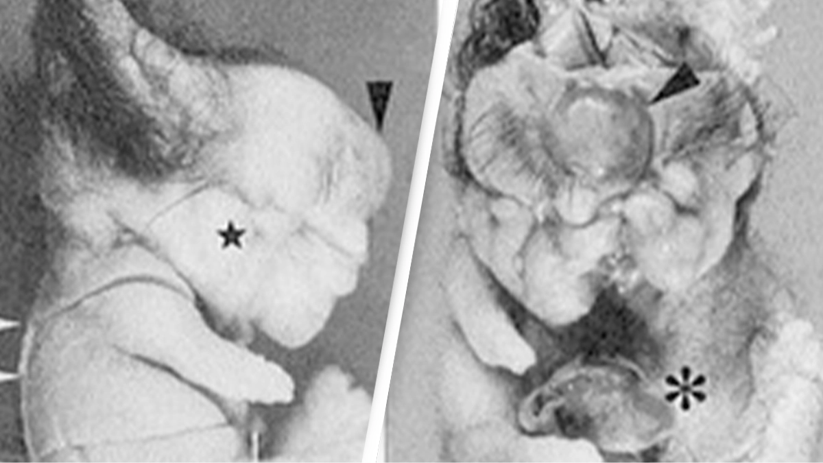
Photos of the tumor are unsettling, to say the least - with its 'doll-like' appearance consisting of limbs, teeth and even hair.
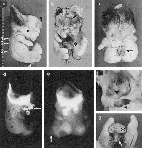
The tumor had a 'doll-like' appearance (Naohiko Kuno, Kenji Kadomatsu, Makoto Nakamura, Takahiko Miwa-Fukuchi, Norio Hirabayashi, Takao Ishizuka)

The abstract for the study read: "A solid mass within the tumor was found to have a head, trunk, and extremities."

The tumor was ultimately diagnosed as a 'mature fetiform teratoma (homunculus)' which, according to Cleveland Clinic, is 'a type of dermoid cyst that consists of living tissue and often resembles a malformed fetus'.

"Brain, eye, spinal nerve, ear, teeth, thyroid gland, bone, bone marrow, gut, trachea, blood vessels, and phallic cavernous tissue were confirmed microscopically," the study added.

The researchers went on to describe its 'distinctive features' that were the 'clear anterior-posterior, ventral-dorsal, and left-right axes, with a spatially well-organized arrangement of the organs'.

Horrifyingly, an eye was located on the front of its head, while it even developed a spinal nerve and spinal bones - 'the thyroid gland was anterior to the trachea, and the gut was deep inside the trunk', the study's authors wrote.

The woman's tumor had developed an eye and brain (Getty Stock Image)

They came to an intriguing conclusion 'that the information necessary for organization of the body plan may be conserved and transmitted, even with parthenogenesis' - a form of asexual reproduction.

The researchers continued: "Mature cystic teratomas of the ovary are mostly benign and do not always attract detailed attention.

"However, precise analyses of such tumors may significantly enhance our understanding of both parthenogenetic and normal human development."

The authors of the study were: Naohiko Kuno, Kenji Kadomatsu, Makoto Nakamura, Takahiko Miwa-Fukuchi, Norio Hirabayashi and Takao Ishizuka.

A teratoma tumor was also discovered last decade in the ovaries of a 16-year-old Japanese woman when she underwent surgery to remove her appendix.

Choose your content:

5 hours ago
6 hours ago
  • Marvin RECINOS / AFP via Getty Images
    5 hours ago

    Director of world’s most dangerous prison reveals why lights are never switched off

    CECOT prison in El Salvador holds 40,000 of the world's most deadly criminals

    News
  • Tennessee Department of Correction
    5 hours ago

    Witnesses recall chilling sounds as death row inmate asked to 'rate his pain' during failed execution

    The Tennessee death row inmate was scheduled to be executed last week

    News
  • Michael Tran/FilmMagic
    5 hours ago

    Matthew Perry's assistant receives prison sentence for his part in actor's tragic death

    Matthew Perry's personal assistant has been sentenced

    News
  • Seros MUYISA / AFP via Getty Images
    6 hours ago

    Ebola outbreak could become 'deadliest on record' as countries the disease has spread to are outlined

    The epidemic is being driven by the rare Bundibugyo virus, which has no proven vaccine

    News
  • Daughter left disturbed after discovering horrific truth of what happened to dad’s body parts that were ‘donated to science’
  • Woman's ultrasound reveals hair and teeth were growing in her uterus for years
  • Woman who had sex with her husband every single day for a year reveals impact it had on her body and relationship
  • Horrifying simulation shows what happened to man who fell into Yellowstone hot spring and was completely dissolved within a day